Abstract
This study aims to present a clinical case and assess the efficacy of the Bimler C Elastic Modeler, a functional orthopedic appliance, in the early treatment of a patient diagnosed with mesiocclusion (mandibular prognathism) based on Bimler and McNamara Cephalometrics. Additionally, we aim to delineate the observed positive changes in facial expression during the course of treatment with this functional orthopedic appliance. The female patient, aged six years and nine months, manifested atypical swallowing, respiratory challenges, and allergic conditions such as rhinitis. Comprehensive examination further revealed facial asymmetry and a smile with lip asymmetry. Intra-oral examination exposed a rightward deviation of the mandible, crossbite, open bite, and mesiocclusion. The proposed intervention encompassed the application of the Bimler Elastic Modeler (BEM) functional orthopedic appliance. The documented treatment duration spanned 12 months, with ongoing monitoring every 2 or 3 months. The treatment, utilizing the BEM functional orthopedic appliance, coupled with exercises and adjustments, improved mandibular and tongue posture, enhancing overall chewing balance. The appliance effectively repositioned the mandible to a more balanced position approaching normocclusion, achieving this without causing pain or discomfort and without the necessity for elastic or constant forces. Given the crucial role of facial expression muscles in these activities, a pronounced enhancement in facial harmony was observed. These affirmative outcomes significantly contributed to heightened patient engagement throughout the treatment process and a concomitant enhancement in patient self-esteem, attributable to the documented aesthetic and functional ameliorations.
1. Introduction
The origins of Jaw Functional Orthopedics trace back to 1881 when Wilhelm Roux delved into the study of the influence of natural forces and functional stimulation [1-3]. In 1949, Bimler introduced three fundamental appliance types – A, B, and C [2-5], leveraging forces derived from muscles, particularly the tongue, to effectively address orofacial muscle dysfunctions [1-4].
Bimler Elastic Modelers (BEM), these appliances operate as a systemic, dynamic, and functional treatment modality [5, 6]. Understanding their mechanism, akin to springs transmitting neural signals throughout the system, requires a grasp of embryology and neurophysiology [6, 7].
Jaw Functional Orthopedics, a specialized field, focuses on diagnosing, preventing, and treating functional and growth issues impacting occlusion and its skeletal foundations. The goal is to eliminate hindrances impeding the proper growth and development of the stomatognathic system (SS) through guidance, education, occlusal adjustments, and intra-oral appliances [6, 8, 9].
Initiating early treatment and monitoring a child’s development are crucial to identify any functional changes [10]. The functions of the SS play a pivotal role in jaw development. Malocclusion can mirror functional imbalances, such as breathing, swallowing, and chewing, influencing the development of skeletal structures, masticatory muscles, and facial expression [11]. Functional orthopedic appliances strive to restore balance to the SS [3, 12, 13].
Even in cases with an unfavorable prognosis, timely diagnosis and interception significantly enhance the success rates of treatment with functional orthopedic appliances [14]. Correcting interferences promptly through functional guidance, occlusal adjustments, and the use of appliances yields better responses, restoring system functionality and ensuring proper growth and development [15, 16]. This study presents a case report classified according to Bimler Cephalometry [6] and Mc Namara, focusing on mesiocclusion (prognathism) and/or anterior crossbite, treated with BEM type C.
2. Case report
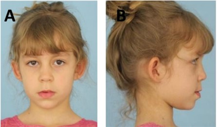
Patient M.V.L.S, a 6-year-and-9-month-old female, presented at a private Jaw Functional Orthopedics dental clinic. After the initial conversation, the Informed Consent Form was presented and signed by those responsible. The anamnesis revealed a normal birth, with the responsible relating the use of a pacifier until six and a half years. Although her overall development was normal, family concerns arose due to a hereditary tendency for enlarged jaws. Noteworthy observations included sporadic chewing of hard foods and a habitual open mouth posture during various activities. The patient demonstrated independent performance of daily tasks. During the extra-oral clinical examination, distinctive findings encompassed atypical swallowing, passive lip sealing, oral breathing, and a history of allergic conditions such as a runny nose. Additionally, facial asymmetry with right-sided deviation was evident (illustrated in Fig. 1).
Fig. 1a) Initial extra-oral photographs of the front and b) right side
The intra-oral clinical examination revealed no physiological wear. The patient displayed an atretic maxilla with a notable deviation of the mandible to the right side and a slight mesiocclusion (Figs. 2(a, b)). McNamara’s analysis identified a mild retrusion of the maxilla and protrusion of the mandible, with smaller maxillary and larger mandibular dimensions according to the table and an increased Afai (linear distance between the points ENA and Me projected perpendicular to the N-perpendicular). Bimler’s analysis indicated an upper mesoprosope and lower leptoprosope pattern, suggesting vertical growth of the maxilla with retroinclination. The Lavergne/Petrovic analysis categorized the patient under rotational growth group P1NOB, with a tissue level 2 growth potential. There was posterior rotation of the mandible, and the mandibular growth potential was smaller than the maxilla, resulting in a favorable prognosis (Figs. 2(c, d)).
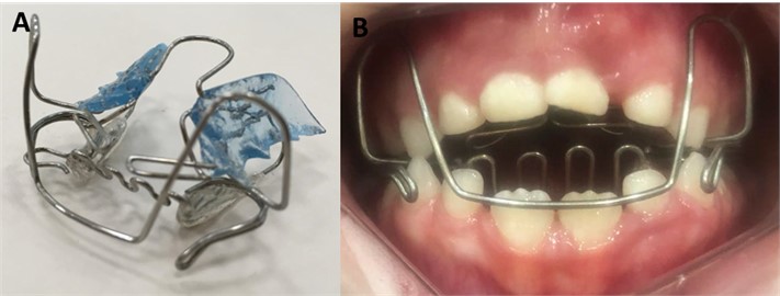
The author systematically evaluated, studied, and diagnosed the presented case in accordance with the principles and regulations governing Jaw Functional Orthopedics [6, 9]. Subsequent to the diagnosis, the proposed treatment plan involved the utilization of the BEM, a functional orthopedic appliance. The appliance, crafted by a skilled orthotechnician, adhered to neurophysiological principles and technical guidelines provided by the author, following the contemporary design of the Bimler appliance (Fig. 3). The treatment objective aimed to gently and progressively move the mandible to a more posterior position without compromising the temporomandibular joint. This repositioning was intended to induce postural changes in a specified Determined Area (DA), fostering a new neural excitation [6-8].
Fig. 2a) Initial intraoral photo in occlusion; b) initial intraoral photo of the palate; c) initial teleradiography; d) initial cephalometry
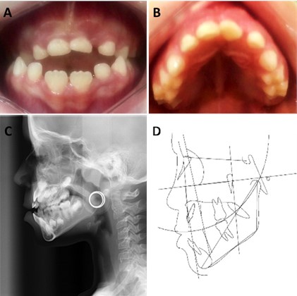
Fig. 3a) Bimler C appliance, b) intra-oral photo after installing the Bimler C appliance
The functional orthopedic apppliance was successfully prescribed, leading to a modification in mandibular posture. Monthly adjustments were performed on the dorsal arch, frontal laces, Eschler arch and Coffin. After 6 months, we observed some initial changes, such as a reduction in mesiocclusion, teeth with space for eruption, and uncrossing of the deciduous canines (Fig. 4(a)). The functional orthopedic treatment spanned approximately 12 months (Fig. 4(b)).
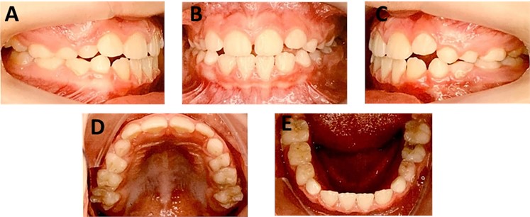
After one year of treatment, the patient showed improvement in the posture of his lips and tongue, in addition to the correction of the malocclusion, evident in the photographs (Fig. 5(a, b)). Additionally, noteworthy improvements in various functions, including breathing, chewing, and swallowing. The reported case demonstrated satisfactory changes, facilitating progress in treatment and fostering a more balanced masticatory function. The appliance effectively repositioned the mandible close to normocclusion, as evidenced by radiography and cephalometry (Fig. 6). Importantly, these improvements were achieved without causing pain or discomfort and without the use of elastic or constant forces [7, 8]. Notably, the patient’s self-esteem and social interaction experienced positive changes, as reported by their parents (Fig. 7).
Fig. 4a) Six months – initial changes, b) 12 months treatment
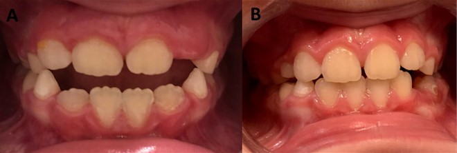
Fig. 5Extraoral photography after 12 months of treatment, frontal, profile on the right side and smile, with changes in lip, facials, muscles and aesthetic
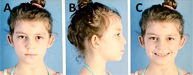
Fig. 6a) Final teleradiography – 12 months, b) final cephalometry – 12 months
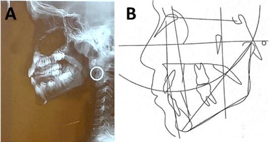
Fig. 7a) Intraoral photography after 12 months of treatment, right lateral; b) frontal; c) left lateral, d) occlusal of the maxilla and e) mandible
3. Conclusions
The study showcased the efficacy of the appliance in successfully attaining correct the installed malocclusion in addition to yields better responses, restoring of SS functionality and ensuring proper growth and development of the maxillary bones. Functional orthopedic appliances emerge as a potentially effective alternative for addressing mesiocclusions, particularly when administered during the early stages of growth and development.
References
-
F. J. Aguila, “Theoretical and Practical Orthodontics,” (in Portuguese), Santos, 2001.
-
T. M. Graber and B. Neumann, Removable Orthodontic Apparatus. (in Spanish), Buenos Aires: Editorial Médica Panamericana, 1979, pp. 337–4801.
-
B. Bimler, “Peter Bimler: uma história de pioneirismo,” (in Portuguese), Revista Dental Press de Ortodontia e Ortopedia Facial, Vol. 9, No. 6, pp. 18–20, Dec. 2004, https://doi.org/10.1590/s1415-54192004000600004
-
N. Wahl, “Orthodontics in 3 millennia. Chapter 2: Entering the modern era,” American Journal of Orthodontics and Dentofacial Orthopedics, Vol. 127, No. 4, pp. 510–515, Apr. 2005, https://doi.org/10.1016/j.ajodo.2005.01.002
-
H. P. Bimler and B. Bimler, “Elastic modelator of Bimler,” Journal Brasileiro de Ortodontia e Ortopedia Maxilar, Vol. 2, No. 7, pp. 57–64, 1997.
-
W. A. Simões, Functional Jaw Orthopedics – Through Neuro-occlusal rehabilitation. (in Portuguese), São Paulo: Artes Médicas, 2003.
-
W. A. Simões, “A study on proprioception and exteroception when Bimler’s, Planas’ and Frankel’s technics are used,” Ortodontia, Vol. 7, No. 2, pp. 153–161, 1974.
-
R. A. Torres, M. H. C. Almeida, T. Wassall, K. J. N. Ferrer, and F. L. C. Bianchini, “Bimler orthopedic functional appliance therapy,” (in Portuguese), R.G.O, Vol. 51, No. 4, pp. 317–322, 2003.
-
G. P. Arceguet, “Etiopathogenic diagnosis in functional maxillary orthopedics,” (in Spanish), Revista de la Asociación Argentina de Ortopedia Funcional de los Maxilares, Vol. 40, No. 1, pp. 9–20, 2014.
-
C. M. C. T. Maranhão, G. R. P. Santos, C. A. S. Sá, M. F. L. Silva, and S. M. S. Silva, “Angle Class III pediatric patient treated with a Fränkel 3 function regulating device,” (in Portuguese), Archives of health investigation, Vol. 7, No. 5, 2018.
-
T. Gedrange and W. Harzer, “Muscle Influence on Postnatal Craniofacial Development and Diagnostics,” Journal of Orofacial Orthopedics, Vol. 65, No. 6, pp. 451–466, Nov. 2004, https://doi.org/10.1007/s00056-004-0405-0
-
R. C. B. Bariani, R. Bigliazzi, M. Cappellette Junior, G. Moreira, and R. R. Fujita, “Effectiveness of functional orthodontic appliances in obstructive sleep apnea treatment in children: literature review,” Brazilian Journal of Otorhinolaryngology, Vol. 88, No. 2, pp. 263–278, Mar. 2022, https://doi.org/10.1016/j.bjorl.2021.02.010
-
M. F. L. D. Silva et al., “Tratamento de mordida aberta anterior em dentadura mista através de aparelho SN6,” (in Portuguese), Research, Society and Development, Vol. 10, No. 13, p. e577101321159, Oct. 2021, https://doi.org/10.33448/rsd-v10i13.21159
-
G. P. L. Silva, D. G. Andrade, T. R. M. Figueiredo, A. D. Lemos, and M. J. A. L. L. A. Arruda, “Angle Class III treatment with reverse maxillary traction and functional jaw orthopedics: case report,” (in Portuguese), Archives of health investigation, 2018.
-
K. E. D. L. Pizzol., C. S. V. Junior, B. A. Almeida, K. B. R. Poquechoque, and V. J. Sanches, “Planas Indirect Tracks: a viable alternative in the early intervention of functional anterior crossbite,” (in Portuguese), Revista Clínica Ortodontia Dental Press, Vol. 17, No. 1, pp. 78–89, 2018, https://doi.org/10.14436/1676-6849.17.1.078-089
-
A. B. Rodrigues, M. M. Uribe, C. A. Morales, and C. H. Martínez-Cajas, “Early treatment of skeletal Class II malocclusions – comparison of three functional appliances: Bionator, Klammt, SN1,” (in Portuguese), Ortodontia SPO, Vol. 46, No. 6, pp. 558–563, 2013.
About this article
The authors have not disclosed any funding.
The datasets generated during and/or analyzed during the current study are available from the corresponding author on reasonable request.
Márcia do Amaral Sampaio: conceptualization; investigation; methodology, writing-original draft preparation; writing-review and editing. Luciano Aparecido de Almeida Junior: conceptualization; investigation; literature review; writing-original draft preparation; writing-review and editing.
The authors declare that they have no conflict of interest.
The Informed Consent Form was presented and signed by those responsible.

