Abstract
This theoretical research is part of the educational and scientific project “Bioelectronic medicine or look at medicine differently” of research work of the Department of Internal Medicine and Emergency Medicine of Poltava State Medical University (23, Shevchenko St., 36011, Poltava, Ukraine) on “Development of algorithms and technologies for implementing a Healthy Lifestyle in patients with Non-Communicable Diseases (NCDs) based on the study of functional status” (state registration number 0121U108237: UDC 613 616-056-06: 616.1 / 9-03). The aim of this theoretical research was to create a systemic medical analysis of the nutrition of the scientific evidence of the Vega Test Method in the complex clinical treatment of patients with Non-Сommunicable Diseases, to determine the current state of health problems and prospects for using the method from the perspective of Complex Medicine. Conclusions: Vega Test Method is a promising modern science-intensive computerized instrumental technique that should be introduced into Clinical Medicine for the examination of patients with NCDs. Existing problems of introducing the Vega Test Method into Clinical Medicine are solved thanks to the progress of fundamental science and Quantum Physics, which leads to a paradigm shift in views on the functioning of the human body. The Vega Test Method has a modern scientific biophysical justification of validity mechanisms based on knowledge of the Magneto-Electrochemical Theory of Metabolism and Life, the Theory of the Electromagnetic Field, and the Concept of Biophoton Signaling. The use of the Vega Test Method in Clinical Medicine for the examination of patients with NCDs is important for the development of Complex Medicine.
Highlights
- A quick look at the VEGATEST-Method with the position of Complex Medicine.
- Vega Test Method is a promising modern science-intensive computerized instrumental technique that should be introduced into Clinical Medicine for the examination of patients with NCDs.
- Existing problems of introducing the Vega Test Method into Clinical Medicine are solved thanks to the progress of fundamental science and Quantum Physics, which leads to a paradigm shift in views on the functioning of the human body.
- The Vega Test Method has a modern scientific biophysical justification of validity mechanisms based on knowledge of the Magneto-Electrochemical Theory of Metabolism and Life, the Theory of the Electromagnetic Field, and the Concept of Biophoton Signaling.
- The use of the Vega Test Method in Clinical Medicine for the examination of patients with NCDs is important for the development of Complex Medicine.
1. Introduction
The term chronic non-communicable diseases (NCDs) unites a group of diseases characterized by a long course, resulting from the influence of a combination of genetic, physiological, environmental and behavioral factors. The main types of NCDs include Cardiovascular Diseases, Malignant Neoplasms, Chronic Respiratory Diseases, Diabetes [1].
In 2014-2024, according to the World Health Organization (WHO), Non-Communicable Diseases (NCDs) annually cause the death of 41 million people, including 15 million people who have not reached the age of old age [1]. NCDs incidence rate has reached a pandemic [1-4]. The spread of NCDs impedes the implementation of the 2030 Agenda for Development, one of which is to reduce by a third the chance of dying from any of the four types of NCDs among people aged 30 to 70 by 2030 [5-7]. NCDs are a major medical and social problem that threatens the achievement of humanity's goals by 2030. Humanity has been trying to solve the problem of NCDs for several decades. In 2019, the WHO World Assembly extended the Global Plan of Action for the Prevention and Control of NCDs 2013-2020 to 2030 and called for the development of a roadmap for the implementation of the Plan for 2023-2030 to accelerate progress in the field of NCD prevention and control. The road map promotes the implementation of measures to achieve a set of nine global goals, allowing to make the maximum contribution to the prevention and treatment of NCDs [8-11]. But so far the set goals have still not been achieved, despite significant progress in medicine and pharmacology. This determines the urgency of continuing the scientific search for ways to increase the effectiveness of prevention and treatment of NCDs in order to overcome this world-class medical and social problem. That is why the future priority of medicine and health care should be systemic medical approaches with the generalization of modern fundamental scientific knowledge as a further basis for deepening the etiopathogenesis of NCDs and finding the latest modern technologies for examining patients with NCDs.
In the 21st century, a characteristic feature of patients has become the presence of several chronic NCDs at the same time. There is an increase in the number of NCD nosologies with the progression of pathology and with increasing age of the patient [12-14]. Polymorbidity/comorbidity has become a distinctive feature of the modern patient. This complicates the clinical work of the doctor. On the one hand, for the correct selection of adequate etiopathogenetic therapy, it is necessary to diagnose all the pathology of the organs and organ systems that the patient has. On the other hand, the prescription of pharmacotherapy for several NCD nosologies is dangerous due to polypharmacy and an increased risk of adverse reactions and side effects of drugs. The presence of multiple NCD pathologies in a patient determines the prescription and performance of a larger number of laboratory tests and instrumental examinations. A chronically ill patient must be prescribed them more times. All this becomes a significant financial burden for the patient and for insurance medicine, etc. Therefore, modern medicine continues to face the challenge of finding new diagnostic approaches for a quick, non-invasive, financially accessible, valid pre-screening examination of multimorbid patients with NCDs in the office of a family medicine doctor or other therapeutic specialist (cardiologist, nephrologist, gastroenterologist, etc.).
For the practical achievement of the above at primary levels, the issue of introducing modern science-intensive instrumental methods remains relevant. It is this that can contribute to the identification and stratification of the latest risk factors for NCDs, provide an opportunity for an objective quantitative assessment of the patient's health level and create conditions for the prevention of NCDs, control over the effectiveness of healthy lifestyle measures, and the possibility of applying personalized approaches to patients in accordance with promising modern global health care models, in particular 4P-medicine /P4 medicine. The use of such methods should contribute to the optimization of prediction, prevention, personalized approach, and participation in the practical activities of modern therapeutic doctors [15, 16].
In order to optimize the examination and management of patients with NCDs in the course of a scientific study aimed at solving the problem of NCDs, a fragment of a theoretical study was performed to find modern science-intensive instrumental methods that could improve the diagnostic process during the general clinical examination of a patient with NCDs. On the basis of the performed system analysis, it was established that the vegetative resonance testing technique meets the requirements of scientific research and is promising for the indicated use. The aim of this theoretical study was to carry out a systematic medical analysis of the issues of the scientific validity of using the Vega Test Method in the complex clinical examination of patients with NCDs, to summarize modern views on the problem and prospects of using the method from the point of view of Complex Medicine.
2. Materials and methods
This theoretical research is part of the educational and scientific project “Bioelectronic medicine or look at medicine differently” of research work of the Department of Internal Medicine and Emergency Medicine of Poltava State Medical University (23, Shevchenko St., 36011, Poltava, Ukraine) on “Development of algorithms and technologies for implementing a Healthy Lifestyle in patients with Non-Communicable Diseases (NCDs) based on the study of functional status” (state registration number 0121U108237: UDC 613 616-056-06: 616.1 / 9-03). This theoretical study was carried out by an international interdisciplinary team under scientific cooperation agreements.
General scientific methods of system analysis were used (logical methods, method of constructing theory, dismemberment and integration of elements of the studied system, logical, historical research, imaginary experiment, analysis, induction, deduction and synthesis of knowledge, and rules of normative nature).
3. Results and discussion
A systematic medical analysis was carried out regarding the scientific validity of the use of Vega Test Method in the comprehensive clinical examination of patients with NCDs. A generalization of modern views on the biophysical substantiation of the method's capabilities, its problems and prospects are given in the relevant subsections of the review.
3.1. Aspects of the biophysical substantiation of the Vega test method as a modern science-intensive instrumental technique
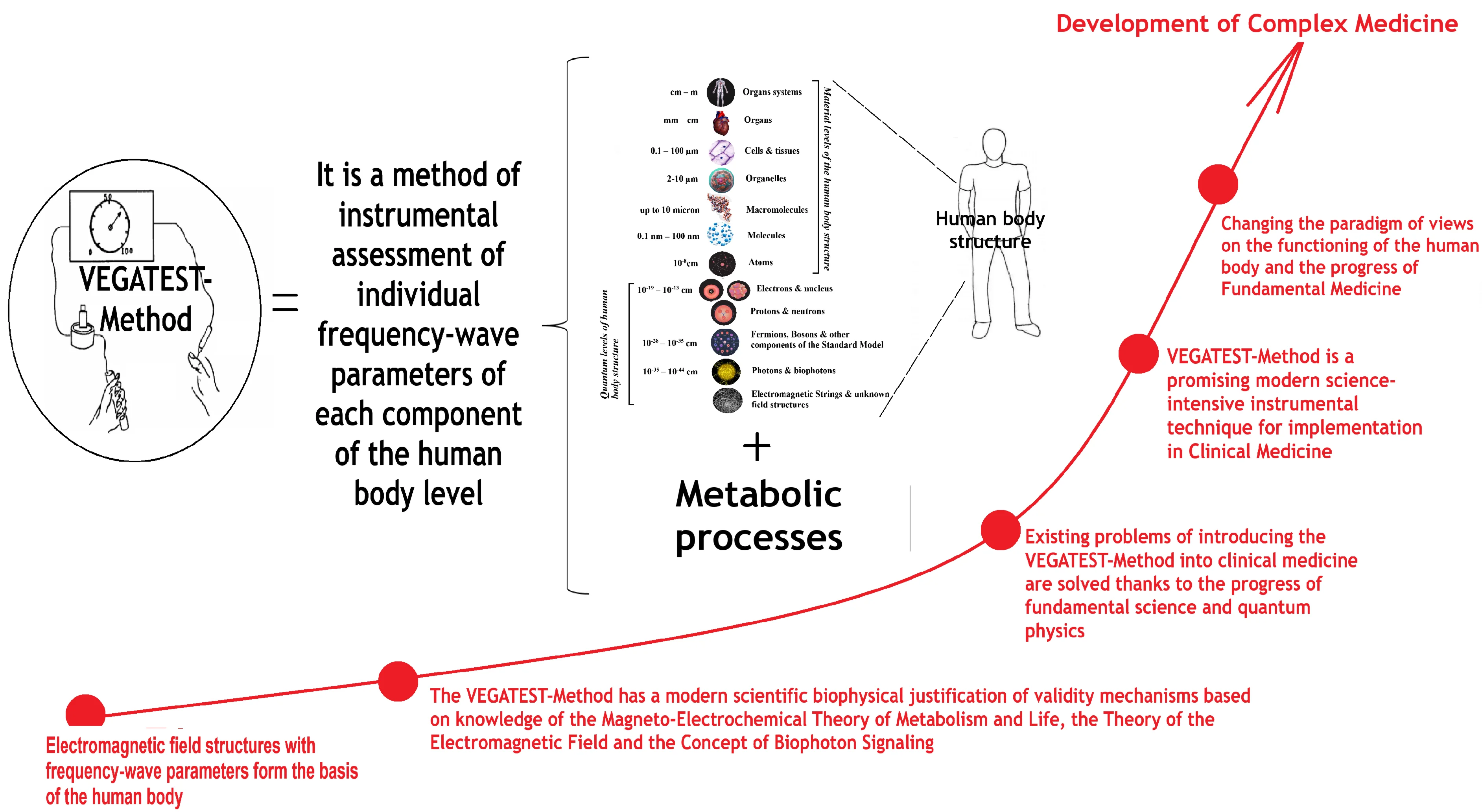
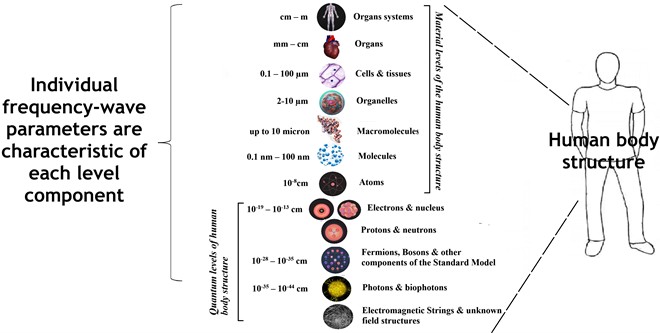
The modern paradigm of the structure of the atoms of the human body according to the Standard Model constitutes the fundamental basis of the biophysical justification of the use of Vega Test Method. According to the standard structural model, atoms are formed from fermions and bosons, which are electromagnetic field structures, that is, energy [17-20]. The human body is made of atoms. Therefore, at the subatomic level, the human body is formed by fermions and bosons, i.e. electromagnetic field structures/energy. At the quantum level of the body structure, everything is electromagnetic field structures/energy that has different quantum-mechanical characteristics, differs in frequency-wave characteristics and is described in the Standard Model. Also, a number of scientists use string theory when describing the structure of substances, which is also a form of reflection of the state of electromagnetic field structures [21,22]. In the human body, material levels of structure (from atoms to a whole organism) and quantum levels (levels of subatomic structures) can be conventionally distinguished (Fig. 1).
This division is rather arbitrary. This is so because quantum levels are also material levels and objectively exist, although they are not available for free perception by human sense organs. The fact of confirming the existence and functioning of quantum levels in the human body is that all atoms, molecules, cells and, accordingly, tissues and organs formed by them, have their own quantum-mechanical biophysical parameters. In addition to the mass, features of the chemical and morphological structure, each material component of the body (atom, molecule, cell, etc.) has frequency wave characteristics, i.e. emits a specific characteristic electromagnetic signal. And it can technically be registered.
This electromagnetic signal is usually evaluated according to such parameters as the frequency of radiation (Hertz, Hz), the nature of the electromagnetic signal (for example, it is coherent or incoherent), the form of the electromagnetic signal (it is harmonic or inharmonic, simple or complex, etc.), spectral density of energy (power of electromagnetic energy per unit of frequency band, W/Hz), spectral structure of the signal (set of harmonics), total power of the signal, width of the frequency band given by this signal, etc. The specified parameters can be used in diagnostics and determine the presence of chemical substances, biological molecules, cell types, etc. in the human body in vivo. This is the direction of studying the electromagnetic mechanisms of communication of the cells of the human body [23-28].
Fig. 1Graphic representation of the structural levels of organization of the human body, taking into account modern fundamental biophysical knowledge
Chemical interaction between atoms occurs on the basis of existing quantum-mechanical characteristics of subatomic structures, which determines their chemical reactivity. That is, all chemical reactions in the human body depend on the electromagnetic state of subatomic structures and arise as a result of its change [29-32]. This determines the fact that all chemical reactions of metabolism in the cells of the human body are a secondary consequence of a change in the electromagnetic state of field structures at the subatomic level of the human body in vivo. This explains the scientific fact that all metabolic processes also have corresponding frequency-wave characteristics. Therefore, metabolic processes in the human body in vivo can be identified by their frequency characteristics with the help of special equipment and methods as well.
Thus, it is clear that every material particle (atom, molecule, biopolymer, cell, tissue, organ, human organism) must now be considered according to the universal law of frequency-wave dualism and as a wave process. Each material part of the human body has frequency-wave parameters of radiation. Complex structures (molecule, biopolymer, cell, tissue, organ, human body) are a total conglomerate of the frequencies of the atoms that make them up. That is, each substance (for example, a vitamin, hormone, amino acid) or type of cells (for example, a virus, bacterium, a cell of the human body) has its own specific constant frequencies of functioning. All material functional levels of the structure of the human body (tissues, organs, organism) can be described in a frequency-wave model, because they are an effective frequency conglomerate of the electromagnetic field due to the summation of the frequencies of all the components that make them up and are present in them [29-32]. This can be used in practice for the purpose of in vivo diagnostics. This scientific paradigm is a biophysical rationale for two diagnostic techniques: nuclear magnetic resonance and vegetative resonance testing.
Each atom has a specific precession frequency of its nucleus. According to this parameter, it is possible to determine the composition of the human body, the presence of certain substances, types of cells and tissues, processes in it. The precession of the nuclei of atoms can be established during the irradiation of the nuclei of atoms with radio waves. It is necessary to constantly change the frequency until it coincides with the frequency of precession of the nuclei. When the precession frequency coincides with the diagnostic frequency, then a resonance occurs, which will be recorded by the measuring device. In this way, the frequency parameters of chemical substances, tissues and processes of functioning of human body organs, most known microorganisms were scientifically established [33-35].
The frequency parameters of human body components continue to be studied. All components of different hierarchical levels of the human body structure have specific frequency-wave parameters. The frequency parameters of large objects, for example, organs and parts of the human body, are studied using the resonance of infrasonic waves. Using resonant infrasonic waves for diagnostics, honest resonances have been established for parts of the human body [36]: for the entire human body in a sitting position, the resonance is 4-6 Hz; resonance for the head in a sitting position with vertical vibrations is in the zone between 20-30 Hz, with horizontal vibrations – 1.5-2 Hz; there is resonance of the eyeballs in the frequency range between 60 and 90 Hz; for organs located in the chest and abdominal cavity, frequencies of 3-3.5 Hz are resonant. For example, the resonance of the heart is 1-2 Hz; the resonance of the stomach is 2-3 Hz; intestinal resonance is 2-4 Hz; the kidney resonance is 6-8 Hz, etc. For micro-objects, resonant frequencies can be calculated. For example, rotational resonances were calculated for deoxyribonucleic acid molecules (DNA) [36]: Y chromosome is 4.00 Hz; The X chromosome is 2.46 Hz; the first chromosome is 1.91 Hz; the second chromosome is 1.97 Hz; the third chromosome is 2.19 Hz; the fourth chromosome is 2.22 Hz; the fifth chromosome is 2.29 Hz; the sixth chromosome is 2.37 Hz; the seventh chromosome is 2.44 Hz; the eighth chromosome has 2.54 Hz; the ninth chromosome is 2.59 Hz; the tenth chromosome is 2.64 Hz; the eleventh chromosome is 2.65 Hz; the twelfth chromosome is 2.66 Hz; the thirteenth chromosome is 2.87 Hz; the fourteenth chromosome is 2.97 Hz; the fifteenth chromosome is 3.04 Hz; the sixteenth chromosome is 3.34 Hz; the seventeenth chromosome is 3.41 Hz; the eighteenth chromosome is 3.48 Hz; the nineteenth chromosome is 4.00 Hz; the twentieth chromosome is 3.87 Hz; the twenty-first chromosome is 4.43 Hz; the twenty-second chromosome is 4.29 Hz. Currently, the frequency spectrum of substances and processes of the human body has been sufficiently studied. Available information bases with general access to them. For example, The Consolidated Annotated Frequency List - CAFL [37], The Non-Consolidated Frequency List - NCFL, The All-Frequencies CAFL (AFCAFL) [38], etc. Official electronic and paper editions are published [35].
The principle of resonance is the basis of the Method of Nuclear Magnetic Resonance. The purpose of the Method of Nuclear Magnetic Resonance is to investigate the complete composition of tissues. For this, it is necessary to create a sufficiently powerful electromagnetic field to affect the precession of each atomic nucleus of the structure of the human body, obtain a resonant response from it, and based on the summation of these resonances, graphically display the composition of a section of the human body. Edward Purcell and Felix Bloch (USA) received the Nobel Prize in Physics in 1952 for the discovery of the phenomenon of resonance. Peter Mansfield (Great Britain) and Paul Lauterbur (USA) received the Nobel Prize in Physiology and Medicine in 2003 for the development of a diagnostic Method of Magnetic Resonance Imaging [39, 40].
In contrast to the Method of Magnetic Nuclear Resonance, Vega Test Method is a method that registers exactly the resonant response to the application of only the test diagnostic frequency or complex of frequencies. The method does not investigate the state of precession of all the nuclei of the body's atoms, unlike the Method of Nuclear Magnetic Resonance. Therefore, there is no need to create powerful electromagnetic fields for external electromagnetic influence on the human body, as when performing the Method of Nuclear Magnetic Resonance. Vega Test Method has a different clinical task than Method of Nuclear Magnetic Resonance. Vega Test Method is designed to check the presence of frequencies of specific substances, cells and processes in the body. For this, it is not necessary to irradiate the whole body with a powerful electromagnetic field, but it is enough to apply test frequencies on a weak electrical signal of 3 mA locally to one biologically active point through a diagnostic probe and record the response. The Vega Test Method or Vegetative Resonance test was developed in the early 70s of the 20th century by the German doctor Helmut Schimmel. Scientific studies of the Schimmel method took more than 15 years. Vega Test Method (VEGATEST-Method or Vegetative Reflex Test, VRT) is the culmination of approximately thirty years of development and experience of German Electroacupuncture. Schimmel optimized the Electropuncture Diagnostic Methods of R. Voll (Elektroakupunktur nach Voll, EAV) and Bioelectronic Functional Diagnostics V. Schmidt and H. Pflaum. Thanks to Schimmel's method and technology, it became possible to use one biologically active measurement point on the patient's hand for diagnosis, and a unique opportunity appeared to reconstruct etiopathogenetic chains from frequency-wave drug complexes during dermatoelectropuncture. It has become more convenient and more informative for the practical use of the dermatoelectropuncture technique by doctors [41]. But the biophysical mechanism of this phenomenon remained unclear. This gave rise to scientific discussions and stopped the wide spread of the method. In 2019, scientific work was started on the systematization of modern biophysical knowledge with its further extrapolation to medical knowledge. Work was started on the conceptualization of the Magnetoelectrochemical Theory of Metabolism and Life [29-32]. In 2024, a working Concept of Biophoton Signaling was developed within the educational and medical project “Bioelectronic medicine or look at medicine differently” [42]. The Concept of Biophoton Signaling is a theoretical framework that explains the biophysical mechanisms of intracellular communication and transfer of energy and information between different cells, tissues and body segments. This conceptualized modern ideas about the role of biophotons in the implementation of reflex connections between organs through energy channels in the human body, which are defined in Traditional Medicine as meridians. This direction of theoretical research was carried out in accordance with the position of WHO, namely the concept of conclusions “Traditional Medicine Global Summit 2023 meeting report: Gujarat Declaration” [43]. According to [43], research into the mechanisms of effectiveness of Traditional Medicine is relevant in modern science. The Concept of Biophoton Signaling [42] explains the mechanism of how information about the state of the entire human body becomes available for registration at biological points of the body. This happens thanks to the biophotonic mechanisms of cell communication based on the Electromagnetic Field Theory. The general essence of the Concept of Biophotonic Signaling is presented in Fig. 2.
Each cell generates its electromagnetic signal due to the electromagnetic activity of the biopolymers of its membrane structures [44]. This electromagnetic generation occurs due to the oscillations of biopolymers with anhydride groups [44]. Molecules of adenosine triphosphate (ATP), which are carriers of incoherent energy, enter these biopolymers. Biopolymers convert this incoherent energy into coherent energy in the form of solitons. Solitons are a form of energy in the form of a standing wave, which is able to transfer energy and information without loss [45-47]. In this way, biopolomers create an energy flow between themselves and transfer it to the systems of structured energy-stressed liquid crystal structures of body water [48, 49]. Biopolymer energy is converted into an electromagnetic coherent state by its own oscillations [50-53]. But where do they get the information component from? Information to them comes from DNA. The electromagnetic mechanisms of DNA functioning cause the transmission of information in the form of biophotons from the deoxyribonucleic acids of the cell nucleus and mitochondria [54-56]. It has been proven that mitochondria play a key role in the generation of biophotons [42], in the metabolism and pathogenesis of NCDs [57, 58]. Biophotons are quanta of energy of the electromagnetic field, which are also in the state of solitons and are an electromagnetic means of communication between the nucleus, mitochondria and other compartments of the cell [59-65]. Biophoton mechanisms ensure the translation of information about all metabolic processes of the cell [66-70] and this information falls on the energy generated by polymers and is further translated on it according to the principles of the Field Theory [71]. Therefore, Biophotonics of the human body is a promising scientific direction now [72-74]. But how does this information become available at the same time in every biological point of the human body? The electromagnetic signal in free space should be attenuated. But, first of all, an electromagnetic signal in the form of solitons can be transmitted through the water liquid crystal energy-stressed structures of the cell matrix without loss, since the molecules of structured water are semiconductors [48]. Secondly, this signal can be transmitted without loss through lipoprotein liquid crystal structures of lipid layers of membranes [29-32]. Third, this signal can be transmitted without loss through the connective tissue, which also has quantum mechanical structural features that allow it to be a semiconductor [75, 76]. At the level of the organism, these are myofascial connections or “Muscle Trains” [77-79]. They transmit energy between body segments. They topographically correspond to the external meridians of ancient Eastern medicine and biologically active points of the body are located on them [80-84]. The Primary Vascular System (PVS) communication system complements this information transmission path. This newest anatomical structure was first discovered by the Korean scientist B.H. Kim in 1962 [85-93]. In the 21st century, the existence of PVS was re-proved in accordance with the requirements of modern evidentiary science [94-106]. Now the morphological structures of PVS are described in detail [94-111]. PVS is described as a "fiber-optic" channel system for electromagnetic biophoton signal transmission and knowledge about it needs to be integrated into medical science [112-115]. Now, thanks to the development of the concept of biophoton signaling, the role and place of the PVS in the generation and transmission of energy in the human body can be logically explained. This knowledge was able to be integrated into orthodox medicine. This is a step towards achieving the goals set in 2023 by the WHO at the “Traditional Medicine Global Summit 2023 meeting report: Gujarat Declaration” [43]. According to the quantum field theory, all information in the form of solitons “merges” in the body’s single electromagnetic field, which is formed by the electromagnetic generation of its cells and constantly circulates in the intercellular matrix, in the body's connective tissue system, and in the PVS. In this way, the supply of a test electromagnetic signal with a given frequency makes it possible to carry out diagnostics based on the presence or absence of resonance. This is the key idea of the modern scientific paradigm, which substantiates the biophysical mechanisms of the validity of the vegetative resonance test.
Fig. 2Scheme of energy metabolism in the human body shows the participation of biophotonic signaling. Note: E is the incoherent energy of adenosine triphosphate (ATP). E' is the coherent energy that is formed by the biopolymer. The dotted line on the biopolymer indicates the oscillatory processes of the biopolymer. Stage A is the entry of energy in the form of food into the human body. Stage B is the digestion of food, the assimilation of food substrates, their entry into the blood and cells of the human body. Stage C corresponds to the processes of Ultraweak Photon Emission (UPE) and tissue respiration in the cell, which lead to the formation of biophotons and the ATP molecule, respectively. Stage D occurs on membrane biopolymers (mainly striated muscles) and ensures the transformation of the biochemical energy of the ATP molecule into electromagnetic energy. Stage E is the redistribution and transport of electromagnetic energy from muscles to other parts of the human body along myofascial connections (tendon meridians) and PVS [42]
![Scheme of energy metabolism in the human body shows the participation of biophotonic signaling. Note: E is the incoherent energy of adenosine triphosphate (ATP). E' is the coherent energy that is formed by the biopolymer. The dotted line on the biopolymer indicates the oscillatory processes of the biopolymer. Stage A is the entry of energy in the form of food into the human body. Stage B is the digestion of food, the assimilation of food substrates, their entry into the blood and cells of the human body. Stage C corresponds to the processes of Ultraweak Photon Emission (UPE) and tissue respiration in the cell, which lead to the formation of biophotons and the ATP molecule, respectively. Stage D occurs on membrane biopolymers (mainly striated muscles) and ensures the transformation of the biochemical energy of the ATP molecule into electromagnetic energy. Stage E is the redistribution and transport of electromagnetic energy from muscles to other parts of the human body along myofascial connections (tendon meridians) and PVS [42]](https://static-01.extrica.com/articles/24727/24727-img2.jpg)
3.2. Vega test method as a promising modern science-intensive instrumental technique for introduction into clinical medicine
From 2019-2022, the method of Vega Test Method was studied based on the educational and practical center of Biophotonics and Valeology of the Department of Internal Diseases and Emergency Medicine of the Poltava State Medical University (Ukraine). The following generalization was made based on the theoretical and clinical studies conducted.
The use of Vega Test Method in the clinical practice of doctors of a therapeutic profile opens opportunities for the introduction of modern science-intensive instrumental methods in the clinic of internal medicine when examining patients with NCDs. The use of Vega Test Method in the form of a device to improve the direct work of the doctor with the patient will contribute to the personalization of patient management, rapid pre-screening of existing pathological processes, pathogens, deviations from the norm of biochemical markers, assessment of health and stress levels, selection of adequate individually suitable pharmacological drugs and nutrients needs, etc. Therefore, the development, use and wide implementation of the Vega Test Method in the clinical activity of therapeutic profile doctors/family doctors is promising and will have significant clinical significance in the future. Now, thanks to the presence of certified modern hardware and software complexes, the Vega Test Method is a combination of clinically significant and technically accessible diagnostic digital technology, which is advisable to use during the clinical examination of patients to identify their current functional state. This method makes it possible to carry out an objective assessment of the biophysical frequency-wave parameters of the patient’s body and to use their analysis in clinical practice in the diagnostic and therapeutic aspect. It should be emphasized that if we know the normal functioning frequency of an organ, then if we diagnose its frequency deviation, we can also use biophysical methods in complex treatment to normalize them. This was justified in [116]. In the dynamics of the treatment and after the treatment, the doctor can monitor the effectiveness of the implemented complex therapy based on the frequency spectrum of the functioning of the organs. Thus, the application of the Vega Test Method in therapeutic practice during the objective examination of the patient and in the dynamics of his management enables the doctor to objectively determine the level of his health, to carry out dynamic observation, as well as comprehensive treatment.
Until recently, only a survey was the only method of assessing the patient’s psycho-emotional component and detecting increased levels of stress and anxiety. The study of the information component of the frequency characteristics of cardiac activity confirmed the fact that the rhythmic-wave processes of the human body carry information about the functional state of the body [117, 118]. The vegetative test method is a modern instrumental method that allows you to objectively diagnose the existing stress in the patient. The examination methodology allows, with competent application, to reveal a detailed picture of psycho-emotional disorders and to find an individual adequate treatment approach for their correction, including the use of treatment methods of frequency-wave bioelectronic therapy. This will be detailed in future posts.
Today, the Vega Test Method can be considered as a priority for use by family doctors/doctors of a therapeutic profile. This is so, because autonomic testing is the only method that allows at the first and second levels of providing therapeutic care to perform a comprehensive examination of the patient and establish the main trends of the patient's pathological process already during the initial clinical examination. The method allows you to determine the expected deviations according to all the desired biochemical markers (we are talking about the study of frequency equivalents of biochemical indicators), to objectively investigate the parameters of the patient's psycho-emotional, endocrine and immune status, to determine potentially significant infectious agents for the patient, deficiencies of vitamins, trace elements, amino acids, enzymes and predisposition to the occurrence of oncological pathology [40, 116]. In the daily practical activity of the doctor, this method provides an opportunity to deepen the approaches in the implementation of a rapid comprehensive examination and treatment of the patient according to such objective parameters as the body's resonant biophysical response to external diagnostic frequency signals. The results of the autonomic test can fundamentally increase clinical information about the state of functioning of the patient’s body and open up the latest objectively substantiated possibilities of using a personalized pharmacological approach to the correction of the existing pathology. At the same time, the discussion with the patient of the obtained results with a visual demonstration and explanation of the diagnosed deviations is another tool with which the doctor can additionally motivate the patient to change his lifestyle and conscious attitude to the treatment process. This gives the doctor an additional opportunity to effectively implement the principle of participation in working with the patient. Thus, the application of the vegetative test method also corresponds to the ideas of the 4P-medicine model [15, 16], which is an important guarantee of the quality and effectiveness of providing therapeutic care.
3.3. Problems of introducing the Vega test method into clinical medicine
Despite such significant clinical benefits of its use, the Vega Test Method is used in a rather limited way. It is mostly used by doctors of Alternative Medicine: homeopaths, nutritionists, doctors of scholastic medicine. It is used by some dentists to diagnose the compatibility of dental materials. In the practice of family medicine doctors and doctors of a therapeutic profile (therapists, nephrologists, cardiologists, pulmonologists, allergists, pediatricians, etc.), this method has not yet been implemented. To date, methodological approaches to its use in the clinic of diseases of internal organs have not been developed. And this remains a significant fundamental obstacle that slows down the advancement of the method.
The lack of a methodology for using the Vega Test Method in therapeutic practice results in the lack of an educational process related to the use of this method. Access to mastering the practical skills of this technique is quite limited. So far, the technique is taught only in alternative medicine training institutions.
General problems for this technique are the issue of standardization of the terminology of Alternative medicine and orthodox medicine with the creation of appropriate adapted glossaries. Since the technique was created and developed by scientists-engineers and doctors of Alternative Medicine [41,119], particularly homeopaths, the hardware and software and frequency catalogs operate with a significant number of homeopathic terms and use concepts of Alternative Medicine. It is necessary to carry out further scientific work on the adaptation and “approaching” of the terminology to the requirements of Orthodox Medicine.
A separate debatable problem is the question of the repeatability of the results and their dependence on the physical and psycho-emotional state of the patient and the diagnostician. The question of researching the quantum levels of the human body and the features of energy and information transmission at the nanolevel and deeper (< 1 nm) in the human body are at the beginning of their scientific mastery. Therefore, there are certainly more questions now than scientifically based answers to them. However, it is now clear that the functional and, accordingly, the electromagnetic state of the human body changes every fraction of a second [117]. Therefore, a number of indicators may change rapidly. These questions require additional scientific study and in-depth clinical and experimental research.
In general, we can say that for a long time the Vega Test Method was outside the attention of orthodox science. This created a significant layer of debatable issues and scientifically unfounded remarks around him. Now in the 21st century, against the background of the significant development of fundamental sciences and the development of quantum physics, a fundamentally new scientific paradigm is emerging in the understanding of the electromagnetic foundations of the functioning of the human body at the nanolevel [29-32]. This changes the scientific view of this method and opens a promising way for its practical understanding as a modern science-intensive technology. The latest biophysical knowledge about the role of electromagnetic processes in the phenomenon of life and the phenomenon of the health of the human body allows a fundamentally new perspective on the essence of the vegetative resonance testing method.
3.4. Prospects for introducing the Vega test method into clinical medicine
Currently, as part of the educational and scientific project “Bioelectronic medicine or look at medicine differently”, scientific work continues on the adaptation of the methodology of the Vega Test Method, developed for convenient clinical use by doctors of family medicine and doctors of a therapeutic profile (therapists, nephrologists, cardiologists, pulmonologists, allergists, pediatricians etc.). At the same time, it is proposed to use the Vega Test Method as a promising additional device-improvement to the general clinical examination.
The use of hardware and software complexes for Vega Test Method can allow a transition to a qualitatively new level of objective examination of a patient by a doctor. This will provide the doctor with the opportunity for a comprehensive examination of the patient during the initial clinical examination in the necessary volume: from a complete comprehensive diagnostic examination lasting up to 1.5-2 hours to a few minutes of determination of the necessary functional parameters at the time of the examination. The use of hardware and software complexes to perform the Vega Test Method can be the next step in the technical improvement of the direct work of a doctor with a patient, which at one time became a phonendoscope and a tonometer, a thermometer and an electrocardiograph. It is certain that the obtained data can be used in the work of doctors at the first and second stages of providing therapeutic care to assess the functional state of the patient, his level of health, further determine the direction of personalized lifestyle correction for the purpose of prevention of NCDs, preservation of health as the main of personal and social value both in peacetime and wartime.
Currently, the level of significance of insurance medicine and preventive medicine is increasing. Personal awareness of the importance of one's own health is growing among a significant number of patients who are interested in having a healthy, long, quality life and turn to doctors for the purpose of prevention and prevention of diseases of internal organs. Unfortunately, traditional clinical examinations and traditional additional methods of laboratory-instrumental methods of examination are not suitable for detecting pathological abnormalities at the stage of the preclinical stage of the disease. These methods are aimed at diagnosing pronounced morphological pathological changes in organs and biochemical deviations from the norm. Also, the appointment of a large number of examinations of biochemical indicators is expensive and technically difficult to perform. Thus, with the help of routine laboratory tests, it is difficult to simultaneously assess deficiencies of all amino acids, vitamins, minerals, and hormones and conduct a full screening for infectious agents. Therefore, equipping doctors with hardware and software complexes for performing Vega Test Method, creating relevant thematic improvement courses at the post-graduate stage of training doctors is an appropriate and important step for the development of preventive medicine and meeting the social demand for preventive medicine among the conscious population.
4. Conclusions
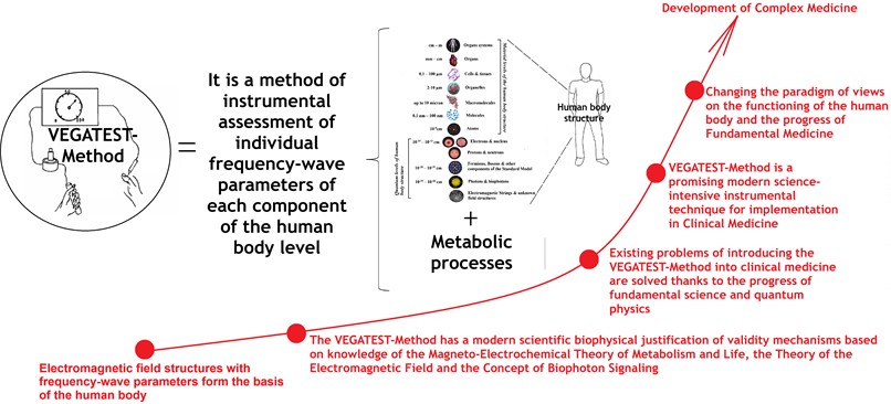
1) Vega Test Method is a promising modern science-intensive computerized instrumental technique that should be introduced into clinical medicine for the examination of patients with NCDs.
2) Existing problems of introducing the Vega Test Method into clinical medicine are solved thanks to the progress of fundamental science and quantum physics, which leads to a paradigm shift in views on the functioning of the human body.
3) The Vega Test Method has a modern scientific biophysical justification of validity mechanisms based on knowledge of the magneto-electrochemical theory of metabolism and life, the theory of the electromagnetic field, and the concept of biophoton signaling.
4) The use of the Vega Test Method in clinical medicine for the examination of patients with NCDs is important for the development of Complex Medicine (Fig. 3).
Fig. 3Graphic abstract. A quick look at the VEGATEST-method with the position of complex medicine
References
-
“Non-communicable diseases fact sheet,” World Health Organization, 2024.
-
“Noncommunicable Diseases,” World Health Organization, 2024.
-
The Lancet, “Non-communicable diseases: What now?,” Lancet, Vol. 399, pp. 1201–1201, 2022, https://doi.org/https://doi.org/info:doi/
-
D. Kostova, P. Richter, G. van Vliet, M. Mahar, and R. L. Moolenaar, “The role of noncommunicable diseases in the pursuit of global health security,” Health Security, Vol. 19, No. 3, pp. 288–301, Jun. 2021, https://doi.org/10.1089/hs.2020.0121
-
J. Kundu and R. Chakraborty, “Socio-economic inequalities in burden of communicable and non-communicable diseases among older adults in India: evidence from longitudinal ageing study in India, 2017-18,” PLOS ONE, Vol. 18, No. 3, p. e0283385, Mar. 2023, https://doi.org/10.1371/journal.pone.0283385
-
C. A. S. Andrade et al., “Inequalities in the burden of non-communicable diseases across European countries: a systematic analysis of the Global Burden of Disease 2019 study,” International Journal for Equity in Health, Vol. 22, No. 1, p. 140, Jul. 2023, https://doi.org/10.1186/s12939-023-01958-8
-
I. R. Hambleton, R. Caixeta, S. M. Jeyaseelan, S. Luciani, and A. J. M. Hennis, “The rising burden of non-communicable diseases in the Americas and the impact of population aging: a secondary analysis of available data,” The Lancet Regional Health – Americas, Vol. 21, p. 100483, May 2023, https://doi.org/10.1016/j.lana.2023.100483
-
“The link between food, nutrition, diet and non-communicable diseases,” NCD Alliance, 2014.
-
J. E. Bennett et al., “NCD countdown 2030: pathways to achieving sustainable development goal target 3.4,” The Lancet, Vol. 396, No. 10255, pp. 918–934, Sep. 2020, https://doi.org/10.1016/s0140-6736(20)31761-x
-
L. Gassner, I. Zechmeister-Koss, and I. Reinsperger, “National strategies for preventing and managing non-communicable diseases in selected countries,” Frontiers in Public Health, Vol. 10, Feb. 2022, https://doi.org/10.3389/fpubh.2022.838051
-
B. Mikkelsen et al., “Life course approach to prevention and control of non-communicable diseases,” BMJ, Vol. 364, p. l257, Jan. 2019, https://doi.org/10.1136/bmj.l257
-
O. A. Asogwa et al., “Multimorbidity of non-communicable diseases in low-income and middle-income countries: a systematic review and meta-analysis,” BMJ Open, Vol. 12, No. 1, p. e049133, Jan. 2022, https://doi.org/10.1136/bmjopen-2021-049133
-
R. Dunn, E. Clayton, E. Wolverson, and A. Hilton, “Conceptualising comorbidity and multimorbidity in dementia: A scoping review and syndemic framework,” Journal of Multimorbidity and Comorbidity, Vol. 12, Sep. 2022, https://doi.org/10.1177/26335565221128432
-
G. Nevoit and M. Potiazhenko, “Clinical and pathogenetic features of the development of non-communicable diseases depending on the degree of comorbidity, the stage of cardiovascular continum,” Bukovinian Medical Herald, Vol. 26, No. 1 (101), pp. 13–22, May 2022, https://doi.org/10.24061/2413-0737.xxvi.1.101.2022.2
-
L.B. Argueta and M.F. Murphy, “4P medicine: prevention, prediction, precision, & participation and its impact in clinical development,” Worldwide Clinical Trials, Jun. 2024.
-
M. Flores, G. Glusman, K. Brogaard, N. D. Price, and L. Hood, “P4 medicine: how systems medicine will transform the healthcare sector and society,” Personalized Medicine, Vol. 10, No. 6, pp. 565–576, Aug. 2013, https://doi.org/10.2217/pme.13.57
-
J. D. Wells, Discovery Beyond the Standard Model of Elementary Particle Physics. Cham: Springer International Publishing, 2020, pp. 1–69, https://doi.org/10.1007/978-3-030-38204-9
-
P. Paganini, Fundamentals of Particle Physics: Understanding the Standard Model. Cambridge University Press, 2023.
-
T. Hübsch, Advanced Concepts in Particle and Field Theory. Cambridge University Press, 2023.
-
V. N. Binhi and A. B. Rubin, “Theoretical Concepts in Magnetobiology after 40 Years of Research,” Cells, Vol. 11, No. 2, p. 274, Jan. 2022, https://doi.org/10.3390/cells11020274
-
B. R. Greene, D. R. Morrison, and J. Polchinski, “String theory,” Proceedings of the National Academy of Sciences of the United States of America, Vol. 95, No. 19, pp. 11039–11040, 1998, https://doi.org/10.1073/pnas.95.19.1103
-
K. Becker, M. Becker, and J. Schwarz, String theory and M-theory: A modern introduction. Cambridge University Press, 2007.
-
M. Levin, G. Pezzulo, and J. M. Finkelstein, “Endogenous bioelectric signaling networks: exploiting voltage gradients for control of growth and form,” Annual Review of Biomedical Engineering, Vol. 19, No. 1, pp. 353–387, Jun. 2017, https://doi.org/10.1146/annurev-bioeng-071114-040647
-
M. Levin, “Bioelectric signaling: Reprogrammable circuits underlying embryogenesis, regeneration, and cancer,” Cell, Vol. 184, No. 8, pp. 1971–1989, Apr. 2021, https://doi.org/10.1016/j.cell.2021.02.034
-
M. Levin, “Endogenous bioelectrical networks store non‐genetic patterning information during development and regeneration,” The Journal of Physiology, Vol. 592, No. 11, pp. 2295–2305, May 2014, https://doi.org/10.1113/jphysiol.2014.271940
-
J. Malmivuo and R. Plonsey, Bioelectromagnetism: Principles and Applications of Bioelectric and Biomagnetic Fields. NY: Oxford University Press, 1995.
-
M. Cifra, J. Z. Fields, and A. Farhadi, “Electromagnetic cellular interactions,” Progress in Biophysics and Molecular Biology, Vol. 105, No. 3, pp. 223–246, May 2011, https://doi.org/10.1016/j.pbiomolbio.2010.07.003
-
R. Vanwijk, “Bio-photons and Bio-communication,” Journal of Scientific Exploration, Vol. 15, No. 2, pp. 183–197, 2001.
-
O. P. Mintser, M. M. Potyazhenko, and G. V. Nevoit, “Magnetoelectrochemical Theory of Metabolism. Volume 1 Conceptualization,” (in Ukrainian), Interservice, Kyiv-Poltava, 2021.
-
O. P. Mintser, M. M. Potiazhenko, and G. V. Nevoit, “Evaluation of the human bioelectromagnetic field in medicine: the development of methodology and prospects are at the present scientific stage,” Wiadomości Lekarskie, Vol. 5, No. II, pp. 1117–1121, 2019, https://doi.org/0.36740/wlek201905231
-
O. P. Mintser, V. V. Semenets, M. Potiazhenko, P. Podpruzhnykov, and G. V. Nevoit, “The study of the electromagnetic component of the human body as a diagnostic indicator in the examination of patients with Non-Communicable diseases: problem statement,” Wiadomości Lekarskie, Vol. 73, No. 6, pp. 1279–1283, Jan. 2020, https://doi.org/10.36740/wlek202006139
-
O. P. Mintser, M. M. Potiazhenko, and G. V. Nevoit, “Informational analytical representations of the magneto-electrochemical theory of life and health,” Journal of Applied Interdisciplinary Research, Vol. 2, pp. 91–98, Jan. 2023, https://doi.org/10.25929/38d5-p759
-
N. Sylver, The Rife Handbook of Frequency Therapy and Holistic Health Hardcover. Desert Gate, 2011.
-
C. Vértesi, The Use of Radiofrequency in the Medicine. Budapest, 2010.
-
H. Eder, Frequency Therapy Cysts and Polyps: Effect and Application of a Long Underestimated Method. (in German), B0BGFGDKJS, 2022.
-
B. L. Ikhlov, “Infrasound, microwaves and disease prevention,” (in Russian), Modern Problems of Science and Education, Vol. 2, 2017.
-
B. Mcinturff, The Electroherbalism Frequency Lists. Lulu.com, 2007.
-
R. P. Stafford, “The Non-Consolidated Frequency List (NCFL),” ElectroHerbalism, 2007.
-
J. A. Koutcher and C. T. Burt, “Principles of nuclear magnetic resonance,” The Journal of Nuclear Medicine, Vol. 25, No. 1, pp. 101–111, 1984.
-
O. Filiunova, G. Nevoit, M. Potyazhenko, and A. Vainoras, “Bioelectronic medicine for sports: justification of biophysical mechanisms and clinical feasibility of use,” Fitoterapia, Vol. 3, No. 3, pp. 73–82, Jan. 2023, https://doi.org/10.32782/2522-9680-2023-3-73
-
H. W. Schimmel and V. Penzer, Functional Medicine. Haug Verlag, 1996.
-
G. Nevoit et al., “Biophotonics and reflexology: conceptualization of the role of biophotonic signaling,” Fitoterapia, Vol. 3, No. 3, pp. 62–78, Jan. 2024, https://doi.org/10.32782/2522-9680-2024-3-62
-
“WHO traditional medicine global summit 2023 meeting report: Gujarat declaration,” Journal of Ayurveda and Integrative Medicine, Vol. 14, No. 5, p. 100821, Sep. 2023, https://doi.org/10.1016/j.jaim.2023.100821
-
G. Nevoit, I. A. Bumblyte, M. Potyazhenko, and O. Minser, “Modern biophysical view of electromagnetic processes of the phenomenon of life of living biological systems as a promising basis for the development of complex medicine: the role of cell membranes,” Journal of Complexity in Health Sciences, Vol. 5, No. 1, pp. 22–34, Jun. 2022, https://doi.org/10.21595/chs.2022.22787
-
H. Bolterauer, “Quantum effects on the Davydov soliton,” in NATO ASI Series, Vol. 243, Boston, MA: Springer US, 1990, pp. 99–107, https://doi.org/10.1007/978-1-4757-9948-4_7
-
“Davydov’s Soliton revisited: self-trapping of vibrational energy in protein,” in NATO Science Series B, Springer, 1990.
-
T. Dauxois and M. Peyrard, Physics of Solitons. Cambridge University Press, 2006.
-
G. Nevoit, I. A. Bumblyte, M. Potyazhenko, and O. Minser, “Modern biophysical view of electromagnetic processes of the phenomenon of life of living biological systems as a promising basis for the development of complex medicine: the role of water,” Journal of Complexity in Health Sciences, Vol. 5, No. 2, pp. 45–57, Dec. 2022, https://doi.org/10.21595/chs.2022.23089
-
N. A. Bulienkov and E. A. Zheligovskaya, “System-forming functions of bound water in the mechanism of topochemical reactions of formation of ultrathin layers on water surface,” Biophysics, Vol. 58, No. 1, pp. 1–18, Apr. 2013, https://doi.org/10.1134/s0006350913010041
-
A. S. Davydov, “Solitons and energy transfer along protein molecules,” Journal of Theoretical Biology, Vol. 66, No. 2, pp. 379–387, May 1977, https://doi.org/10.1016/0022-5193(77)90178-3
-
A. S. Davydov, “Quantum theory of muscular contraction,” Biophysics, Vol. 19, pp. 684–691, 1974.
-
A. S. Davydov, Biology and Quantum Mechanics. (in 1982), Oxford: Pergamon Press.
-
A. S. Davydov, “The theory of contraction of proteins under their excitation,” Journal of Theoretical Biology, Vol. 38, No. 3, pp. 559–569, Mar. 1973, https://doi.org/10.1016/0022-5193(73)90256-7
-
G. Nevoit, I. A. Bumblyte, M. Potyazhenko, O. Minser, and A. Vainoras, “Modern biophysical view of electromagnetic processes of the phenomenon of life of living biological systems as a promising basis for the development of complex medicine: the role of biophotons,” Journal of Complexity in Health Sciences, Vol. 6, No. 1, pp. 1–15, Jun. 2023, https://doi.org/10.21595/chs.2023.23443
-
C. Scaletta, F. A. Popp, H. J. Niggli, L. A. Applegate, and Y. Yan, “UV-induced DNA damage and ultraweak photon emission in human fibroblastic skin cells: parameters to trigger intra – and extra-cellular photobiostimulation,” Trends in Photochemistry and Photobiology, Vol. 8, pp. 53–65, 2001.
-
K.-P. Schlebusch, W. Maric-Oehler, and F.-A. Popp, “Biophotonics in the infrared spectral range reveal acupuncture meridian structure of the body,” The Journal of Alternative and Complementary Medicine, Vol. 11, No. 1, pp. 171–173, Feb. 2005, https://doi.org/10.1089/acm.2005.11.171
-
S. E. Alway, H. G. Paez, and C. R. Pitzer, “The role of mitochondria in mediation of skeletal muscle repair,” Muscles, Vol. 2, No. 2, pp. 119–163, Mar. 2023, https://doi.org/10.3390/muscles2020011
-
A. Casanova, A. Wevers, S. Navarro-Ledesma, and L. Pruimboom, “Mitochondria: It is all about energy,” Frontiers in Physiology, Vol. 14, Apr. 2023, https://doi.org/10.3389/fphys.2023.1114231
-
M. Bischof, Biophotons – The Light in Our Cells. (in German), Zweitausendeins, 2008.
-
J. Chang, J. Fisch, and F. A. Popp, Biophotons. Dordrecht: Springer Netherlands, 1998, https://doi.org/10.1007/978-94-017-0928-6
-
M. Cifra, E. van Wijk, H. Koch, S. Bosman, and R. van Wijk, “Spontaneous ultra-weak photon emission from human hands is time dependent,” Radioengineering, Vol. 16, No. 2, p. 15, 2007.
-
M. Kobayashi and H. Inaba, “Photon statistics and correlation analysis of ultraweak light originating from living organisms for extraction of biological information,” Applied Optics, Vol. 39, No. 1, p. 183, Jan. 2000, https://doi.org/10.1364/ao.39.000183
-
M. Kobayashi, T. Iwasa, and M. Tada, “Polychromatic spectral pattern analysis of ultra-weak photon emissions from a human body,” Journal of Photochemistry and Photobiology B: Biology, Vol. 159, pp. 186–190, Jun. 2016, https://doi.org/10.1016/j.jphotobiol.2016.03.037
-
P. Madl, “Biophotonics or the light of life. Lecture series: block I/IV – biophysics in life sciences,” University of Salzburg, 2006.
-
H. J. Niggli, “Biophotons: ultraweak light impulses regulate life processes in aging,” Journal of Gerontology and Geriatric Research, Vol. 3, No. 2, Jan. 2014, https://doi.org/10.4172/2167-7182.1000143
-
F. A. Popp, The Message of Food. (in German), Zweitausendeins, 2005.
-
F. A. Popp, “Coupling of Fröhlich-modes as a basis for biological regulation,” in Herbert Fröhlich, FRS, a Physicist Ahead of His Time: a Centennial Celebration of His Life and Work, University of Liverpool, 2006.
-
F. A. Popp, K. H. Li, and Q. Gu, Recent Advances in Biophoton Research and its Applications. Singapore: World Scientific Publishing, 2011.
-
F. A. Popp, W. Nagl, K. H. Li, W. Scholz, O. Weingärtner, and R. Wolf, “Biophoton emission,” Cell Biophysics, Vol. 6, No. 1, pp. 33–52, Mar. 1984, https://doi.org/10.1007/bf02788579
-
J. N. Tinsley et al., “Direct detection of a single photon by humans,” Nature Communications, Vol. 7, No. 1, pp. 12–17, Jul. 2016, https://doi.org/10.1038/ncomms12172
-
A. Wolski, “Theory of electromagnetic fields,” Cockcroft Institute, University of Liverpool, UK, 2011.
-
R. van Wijk, Light in Shaping Life – Biophotons in Biology and Medicine. Geldermalsen: Meluna Research, 2014.
-
R. van Wijk and X. Shen, Biophotonics: Optical Science and Engineering for the 21st Century. New York, NY: Springer, 2005.
-
R. van Wijk, E. P. A. van Wijk, J. Pang, M. Yang, Y. Yan, and J. Han, “Integrating ultra-weak photon emission analysis in mitochondrial research,” Frontiers in Physiology, Vol. 11, p. 717, Jul. 2020, https://doi.org/10.3389/fphys.2020.00717
-
E. van Wijk, M. Groeneveld, J. van der Greef, and R. van Wijk, “Unusual optical properties of collagen and implications for the primo vascular system,” in The Primo Vascular System, pp. 235–241, Sep. 2011, https://doi.org/10.1007/978-1-4614-0601-3_33
-
M. Yip and P. Madl, “The light of life,” Biophotonics, Vol. 6, pp. 303–311, 2006.
-
O. Guntinas-Lichius et al., “Pathogenesis, diagnosis and therapy of facial synkinesis: A systematic review and clinical practice recommendations by the international head and neck scientific group,” Frontiers in Neurology, Vol. 13, Nov. 2022, https://doi.org/10.3389/fneur.2022.1019554
-
T. W. Myers, Anatomy Trains. Myofascial Meridians for Manual Therapists and Movement Professionals. Elsevier, 2020.
-
J. Whatley, J. Perkins, and C. Samuel, “‘Reflexology: exploring the mechanism of action’,” Complementary Therapies in Clinical Practice, Vol. 48, p. 101606, Aug. 2022, https://doi.org/10.1016/j.ctcp.2022.101606
-
L. Dorfer, M. Moser, K. Spindler, F. Bahr, E. Egarter-Vigl, and G. Dohr, “5200-year-old acupuncture in central Europe?,” Science, Vol. 282, No. 5387, pp. 239–239, Oct. 1998, https://doi.org/10.1126/science.282.5387.239f
-
B. Bordoni and M. Simonelli, “The awareness of the fascial system,” Cureus, Vol. 10, No. 10, Oct. 2018, https://doi.org/10.7759/cureus.3397
-
C. Schnorrenberger, “Morphological foundations of acupuncture: an anatomical nomenclature of acupuncture structures,” Acupuncture in Medicine, Vol. 14, No. 2, pp. 89–103, 1996.
-
C. Schnorrenberger, “An interpretation of fundamental ideographs of Chinese medicine. Erroneous Western translations of basic Chinese medical characters reduce the significance of the German Gerac-studies (‘Modellvorhaben’),” Chweizerische Zeitschrift fur Ganzheitsmedizin, Vol. 17, No. 3, pp. 150–156, 2005.
-
C. Schnorrenberger, “Anatomical roots of Chinese medicine and acupuncture,” Anatomie-Eine Historische Grundlage der Chinesischen Medizin und Akupunktur, Vol. 20, No. 3, pp. 163–171, 2008.
-
S. Fujiwara and S. B. Yu, “Bonghan theory’ morphological studies,” Igaku noAyumi, Vol. 60, pp. 567–577, 1967.
-
B. H. Kim, “Study on the reality of acupuncture meridians,” Journal of Jo Sun Medicine, Vol. 9, pp. 5–13, 1962.
-
B. H. Kim, “On the acupuncture meridian system,” Journal of Jo Sun Medicine, Vol. 90, pp. 6–35, 1963.
-
B. H. Kim, “On the Kyungrak System,” Journal of the Academy of Medical Science of the Democratic People’s Republic of Korea, Vol. 90, pp. 1–41, 1963.
-
B.H. Kim, “On the Kyungrak System”, Forein Language Publishing House, Pyongyang, North Korea, 1964, 41 pages.
-
B. H. Kim, Kyungrak System and Sanal Theory. Pyongyang, North Korea: Medical Science Press, 1965.
-
B. H. Kim, “Sanal and hematopoiesis,” Journal of Jo Sun Medicine, Vol. 108, pp. 1–6, 1965.
-
B. H. Kim, “Sanal theory,” Journal of Jo Sun Medicine, Vol. 108, pp. 39–62, 1965.
-
B. H. Kim, “The Kyungrak system,” Journal of Jo Sun Medicine, Vol. 108, pp. 1–38, 1965.
-
A. M. Hossein, T. Yu-Ying, H. Tao, Z. Yu-Qing, C. Yong-Zhe, and Z. Wei-Bo, “Finding a novel threadlike structure on the intra-abdominal organ surface of small pigs by using in vivo trypan blue staining,” in The Primo Vascular System, pp. 63–69, Sep. 2011, https://doi.org/10.1007/978-1-4614-0601-3_9
-
K. A. Kang, “Chronological review on scientific findings of Bonghan system and primo vascular system,” Advances in Experimental Medicine and Biology, Vol. 923, pp. 301–309, Aug. 2016, https://doi.org/10.1007/978-3-319-38810-6_40
-
K. A. Kang, “Bonghan (primo vascular) system, elucidated by Bong Han Kim: Kim’s findings, later verifications, new findings, and prospective: A review,” Precision and Future Medicine, Vol. 6, No. 2, pp. 117–137, Jun. 2022, https://doi.org/10.23838/pfm.2022.00030
-
J. Kim et al., “Spontaneous ultraweak photon emission during the cell population of culture HeLa cell line,” Journal of Health Science, Vol. 53, No. 4, pp. 481–485, 2007.
-
H.-G. Kim, “Achievements of PVS (Primo Vascular System) research from a historical perspective,” Journal of Acupuncture and Meridian Studies, Vol. 15, No. 1, pp. 50–60, Feb. 2022, https://doi.org/10.51507/j.jams.2022.15.1.50
-
J. Kim, J. Jung, and M. Potroz, “Summary of Bong-Han Kim’s publications,” in The Primo Vascular System, pp. 7–17, Sep. 2011, https://doi.org/10.1007/978-1-4614-0601-3_2
-
M. Kobayashi, D. Kikuchi, and H. Okamura, “Imaging of ultraweak spontaneous photon emission from human body displaying diurnal rhythm,” PLoS ONE, Vol. 4, No. 7, p. e6256, Jul. 2009, https://doi.org/10.1371/journal.pone.0006256
-
J. Kwon, K. Y. Baik, B.-C. Lee, K.-S. Soh, N. J. Lee, and C. J. Kang, “Scanning probe microscopy study of microcells from the organ surface Bonghan corpuscle,” Applied Physics Letters, Vol. 90, No. 17, Apr. 2007, https://doi.org/10.1063/1.2732183
-
B. S. Kwon, C. M. Ha, S. Yu, B.-C. Lee, J. Y. Ro, and S. Hwang, “Microscopic nodes and ducts inside lymphatics and on the surface of internal organs are rich in granulocytes and secretory granules,” Cytokine, Vol. 60, No. 2, pp. 587–592, Nov. 2012, https://doi.org/10.1016/j.cyto.2012.07.016
-
B.-C. Lee et al., “Network of endocardial vessels,” Cardiology, Vol. 118, No. 1, pp. 1–7, Apr. 2011, https://doi.org/10.1159/000323844
-
B.C. Lee et al., “Electron microscopic study of novel threadlike structures on the surfaces of mammalian organs,” Microscopy Research and Technique, Vol. 70, No. 1, pp. 34–43, Oct. 2006, https://doi.org/10.1002/jemt.20383
-
C. Lee, S.-K. Seol, B.-C. Lee, Y.-K. Hong, J.-H. Je, and K.-S. Soh, “Alcian blue staining method to visualize Bonghan threads inside large caliber lymphatic vessels and X-Ray microtomography to reveal their microchannels,” Lymphatic Research and Biology, Vol. 4, No. 4, pp. 181–190, Dec. 2006, https://doi.org/10.1089/lrb.2006.4402
-
S. H. Park, “Bioelectrical study of Bonghan system,” Ph.D. Thesis, Seoul National University, Seoul, 2009.
-
F. Scholkmann, Y. Shen, and P. D. Ryu, “Microscopic detection of a red thread-like structure inside primo vessels and primo nodes from the intestine surface of rats,” Matters, Jan. 2019, https://doi.org/10.5167/uzh-177094
-
K.-S. Soh, K. A. Kang, and Y. H. Ryu, “50 years of Bong-Han theory and 10 years of primo vascular system,” Evidence-Based Complementary and Alternative Medicine, Vol. 2013, pp. 1–12, Jan. 2013, https://doi.org/10.1155/2013/587827
-
K. S. Soh, “Bonghan duct and acupuncture meridian as optical channel of biophoton,” Journal of the Korean Physical Society, Vol. 45, p. 1196, 2004.
-
K.-S. Soh, “Bonghan circulatory system as an extension of acupuncture meridians,” Journal of Acupuncture and Meridian Studies, Vol. 2, No. 2, pp. 93–106, Jun. 2009, https://doi.org/10.1016/s2005-2901(09)60041-8
-
K.-S. Soh, K. A. Kang, and D. K. Harrison, The Primo Vascular System: Its Role in Cancer and Regeneration. New York, NY, USA: Springer, 2011.
-
M. Stefanov, “Critical review and comments on B.H. Kim’s work on the primo vascular system,” Journal of Acupuncture and Meridian Studies, Vol. 5, No. 5, pp. 241–247, Oct. 2012, https://doi.org/10.1016/j.jams.2012.07.008
-
M. Stefanov, “Primo vascular system: before the past, bizarre present and peek after the future,” Journal of Acupuncture and Meridian Studies, Vol. 15, No. 1, pp. 61–73, Feb. 2022, https://doi.org/10.51507/j.jams.2022.15.1.61
-
M. Stefanov, M. Potroz, J. Kim, J. Lim, R. Cha, and M.-H. Nam, “The primo vascular system as a new anatomical system,” Journal of Acupuncture and Meridian Studies, Vol. 6, No. 6, pp. 331–338, Dec. 2013, https://doi.org/10.1016/j.jams.2013.10.001
-
J. Kim and M. Stefanov, “Visualizing the peripheral primo vascular system in mice skin by using the polymer Mercox,” Journal of Pharmacopuncture, Vol. 18, No. 3, pp. 75–79, Sep. 2015, https://doi.org/10.3831/kpi.2015.18.028
-
G. Nevoit, O. Filiunova, M. Potyazhenko, O. Minser, I. A. Bumblyte, and A. Vainoras, “Modern biophysical view of electromagnetic processes of the phenomenon of life of living biological systems as a promising basis for the development of complex medicine: towards the concept of Bioelectronic Medicine,” Journal of Complexity in Health Sciences, Vol. 6, No. 2, pp. 49–66, Dec. 2023, https://doi.org/10.21595/chs.2023.23867
-
O. P. Mintser, M. M. Potyazhenko, and G. V. Nevoit, Systemic Medicine. (in Ukrainian), Kyiv-Poltava: Interservice, 2022.
-
G. V. Nevoit et al., “Electro-photonic emission analysis in patients with noncommunicable diseases: essential hypertension,” World of Medicine and Biology, Vol. 19, No. 83, p. 138, Jan. 2023, https://doi.org/10.26724/2079-8334-2023-1-83-138-143
-
G. A. Ulett, S. Han, and J.-S. Han, “Electroacupuncture: mechanisms and clinical application,” Biological Psychiatry, Vol. 44, No. 2, pp. 129–138, Jul. 1998, https://doi.org/10.1016/s0006-3223(97)00394-6
About this article

The authors have not disclosed any funding.
The datasets generated during and/or analyzed during the current study are available from the corresponding author on reasonable request.
Ganna Nevoit: conceptualization, investigation, visualization, writing-original draft. Olena Filyunova: conceptualization, investigation. Svetlana Danylchenko: conceptualization, investigation. Maksim Potyazhenko: conceptualization, supervision, writing-review and editing. Ozar Mintser: conceptualization, methodology, supervision, writing-review and editing. Inga Arune Bumblyte: conceptualization, data curation, methodology, resources, supervision, writing-review and editing. Alfonsas Vainoras: conceptualization, methodology, project administration, supervision, writing-review and editing.
The authors declare that they have no conflict of interest.
This is not applicable. The manuscript is a fragment of theoretical research.
